Where Is The Cytoskeleton Located In A Animal Cell
The nucleus and cellular organelles are non randomly scattered in the cytoplasm. Indeed, there is a functional and structural internal organisation ruled by several types of proteins bundled in filaments, jointly known as cytoskeleton. These filaments form a dynamic scaffolding distributed through the cytosol, although some of them are plant inside the nucleus.
The give-and-take "cytoskeleton" is a morphological and structural term coming from the early observations of cells at electron microscopy. It may pb to a misunderstanding because the cytoskeleton is not a static scaffold for supporting cell structures. Actually, it is a very plastic structure responsible for cell movement and shape, and for organelle organization and movements. The functional variety of the cytoskeleton is a consequence of its molecular features.
P olymerization and depolymerization. Cytoskeleton filaments are formed by polymerization of repeated proteins that practice not establish chemic bonds between each other, merely they are linked through electric forces. In this way, filaments can be assembled (polymerized) and disassembled (depolymerized) easily and according to the cell needs. Jail cell may form and modify filament scaffolds where they are needed. Proteins that forms cytoskeleton filaments are ever changing betwixt polymerized and costless in the cytosol.
P olarization. Some cytoskeletal filaments are polarized structures, that is, all the protein units in the filament are in the aforementioned orientation. Thus, the two ends of the filament are dissimilar. This arrangement is important for filament growing and for those proteins that move along the filament.
R egulation. Cells take many proteins to control the organization and activity of cytoskeleton filaments. They are tools for manipulating the three dimensional scaffold of cytoskeleton filaments. For example, motor proteins are molecules that use cytoskeletal filaments every bit train rails to transport cargoes (vesicles, organelles, macromolecules) through the cytoplasm. Other proteins are involved in filament polymerization-depolymerization, filament stability, or are intermediaries between filaments and other cell structures.
Cytoskeleton performs an amazing amount of functions in eukaryotic cells. It makes cells to move, establishes the cell shape, makes possible the polarity of some cells, distributes intracellular organelles properly, is responsible for the advice between those organelles, and for exocytosis and endocytosis processes, runs cell division (both mitosis and meiosis), is a proficient scaffold for maintaining intracellular organization, resists mechanical forces, withstands jail cell deformations, and many others. Although some homologous cytoskeletal proteins take been found in prokaryotes, cytoskeleton appears to be invented by eukaryotic cells. The mechanical function of cytoskeleton is particularly useful in animal cells, where no cell wall gives consistency to the jail cell. Without a cytoskeleton, animal cells volition break because plasma membrane is merely a canvas of fatty.
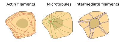
Cytoskeleton is composed of three types of filaments: actin filaments or microfilaments, microtubules, and intermediate filaments (Figure 1). Actin filaments, polymers of repeated units of the actin protein, are in charge of cell movements, endocytosis, phagocytosis, cytokinesis, and other functions. They are besides part of the molecular machinery needed for muscle contraction, and contribute to form some cell junctions (adherent junctions and tight junctions). They are named as microfilaments considering their diameter is lower than those of the other cytoskeleton components. Microtubules, as the name suggests, are tubules made up of dimers of α- and β-tubulin. Microtubules are needed for the intracellular movement of organelles and vesicles, institute the skeleton of cilia and flagella, drive the chromosome segregation during jail cell sectionalisation, etcetera. Actin filaments and microtubules are helped by motor proteins, which are bodily motors that can move along the filaments. Actin filaments and microtubules are used as rails by motor proteins to acquit cargoes. Cargoes may be chromosomes, organelles, or macromolecular complexes. Intermediate filaments are responsible for cell integrity, since they function every bit potent intracellular cables anchored to prison cell junctions like desmosomes and hemidesmosomes. They make possible the adhesion between contiguous cells and cell-extracellular matrix, contributing to the cohesion of tissues. They are specialized in withstanding mechanical forces. Unlike the other components of the cytoskeleton, intermediate filaments are polymers that can be made upward of different families of proteins, such as keratins, vimentins, laminas, and some others.

Source: https://mmegias.webs.uvigo.es/02-english/5-celulas/7-citoesqueleto.php
Posted by: fierropornat.blogspot.com

0 Response to "Where Is The Cytoskeleton Located In A Animal Cell"
Post a Comment